Glaucoma Treatment

Dr. Rudra Prosad Ghosh is an experienced and one of the most trusted Glaucoma specialists in Kolkata. He has done so many glaucoma surgeries during his illustrious career.

Glaucoma damage is permanent—it cannot be reversed. But medicine and surgery help to stop further damage. To treat glaucoma, your ophthalmologist may use one or more of the following treatments.

Medication

Glaucoma is usually controlled with eye drop medicine. Used every day, these eye drops lower eye pressure. Some do this by reducing the amount of aqueous fluid the eye makes. Others reduce pressure by helping fluid flow better through the drainage angle.

Glaucoma medications can help you keep your vision, but they may also produce side effects.

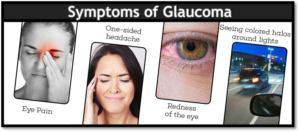
Some eye drops may cause:

  • a stinging or itching sensation
  • red eyes or red skin around the eyes
  • changes in your pulse and heartbeat
  • changes in your energy level
  • changes in breathing (especially if you have asthma or breathing problems)
  • dry mouth
  • blurred vision
  • eyelash growth
  • changes in your eye color, the skin around your eyes or eyelid appearance.

All medications can have side effects. Some drugs can cause problems when taken with other medications. It is important to give your doctor a list of every medicine you take regularly. Be sure to talk with your ophthalmologist if you think you may have side effects from glaucoma medicine.

Never change or stop taking your glaucoma medications without talking to your ophthalmologist. If you are about to run out of your medication, ask your ophthalmologist if you should have your prescription refilled.

Laser Procedure

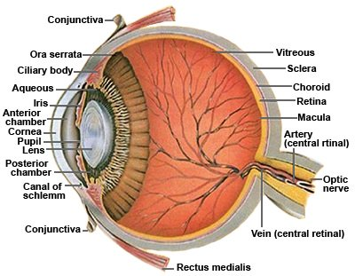
In trabeculectomy, a flap is first created in the sclera (the white part of the eye). Then a small opening is made into the eye to release fluid from the eye.

There are two main types of laser surgery to treat glaucoma. They help aqueous drain from the eye. These procedures are usually done in the ophthalmologist’s office or an outpatient surgery center.

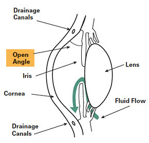
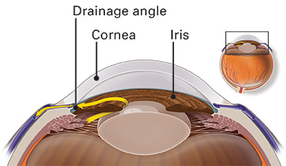
Trabeculoplasty :  This surgery is for people who have open-angle glaucoma. The eye surgeon uses a laser to make the drainage angle work better. That way fluid flows out properly and eye pressure is reduced.

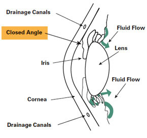
Iridotomy :  This is for people who have angle-closure glaucoma. The ophthalmologist uses a laser to create a tiny hole in the iris. This hole helps fluid flow to the drainage angle.

Operating room surgery

Trabeculectomy. This is where your eye surgeon creates a tiny flap in the sclera. He or she will also create a bubble (like a pocket) in the conjunctiva called a filtration bleb. It is usually hidden under the upper eyelid and cannot be seen. Aqueous humor will be able to drain out of the eye through the flap and into the bleb. In the bleb, the fluid is absorbed by tissue around your eye, lowering eye pressure.

Glaucoma drainage devices. Your ophthalmologist may implant a tiny drainage tube in your eye. The glaucoma drainage implant sends the fluid to a collection area (called a reservoir). Your eye surgeon creates this reservoir beneath the conjunctiva. The fluid is then absorbed into nearby blood vessels.

Your role in glaucoma treatment

Treating glaucoma successfully is a team effort between you and your doctor. Your ophthalmologist will prescribe your glaucoma treatment. It is up to you to follow your doctor’s instructions and use your eye drops.

Once you are taking medications for glaucoma, your ophthalmologist will want to see you regularly. You can expect to visit your ophthalmologist about every 3–6 months. However, this can vary depending on your treatment needs.

What is Glaucoma

A tough white covering called the sclera protects the eye. Part of the white sclera can be seen in the front of the eye. A clear, delicate membrane called the conjunctiva covers the sclera.

At the front of the eye is the cornea. The cornea is the clear part of the eye’s protective covering. It allows light to enter the eye. The iris is the colored part of the eye that contracts and expands so the pupil can let just the right amount of light into the eye. The light is directed by the pupil to the lens. The lens focuses the light onto the retina (lining the back of the eye). Nerve fibers in the retina carry images to the brain through the optic nerve.

Healthy Drainage

The front part of the eye is filled with a clear fluid called intraocular fluid or aqueous humor, made by the ciliary body. The fluid flows out through the pupil. It is then absorbed into the bloodstream through the eye’s drainage system.

This drainage system is a meshwork of drainage canals around the outer edge of the iris. Proper drainage helps keep eye pressure at a normal level. The production, flow, and drainage of this fluid is an active continuous process that is needed for the health of the eye.

The inner pressure of the eye (intraocular pressure or IOP) depends upon the amount of fluid in the eye. If your eye’s drainage system is working properly then fluid can drain out and prevent a buildup. Likewise, if your eye’s fluid system is working properly, then the right amount of fluid will be produced for a healthy eye. Your IOP can vary at different times of the day, but it normally stays within a range that the eye can handle.

The Eye with Glaucoma

In most types of glaucoma, the eye’s drainage system becomes clogged so the intraocular fluid cannot drain. As the fluid builds up, it causes pressure to build within the eye. High pressure damages the

The Optic Disc

You have millions of nerve fibers that run from your retina to the optic nerve. These fibers meet at the optic disc. As fluid pressure within your eye increases, it damages these sensitive nerve fibers and they begin to die. As they die, the disc begins to hollow and develops a cupped or curved shape. If the pressure remains too high for too long, the extra pressure can damage the optic nerve and result in vision loss.

Is There Another Cause?

It was once thought that high intraocular pressure (IOP) was the main cause of this optic nerve damage. Although IOP is clearly a risk factor, we now know that other factors must be involved because people with “normal” IOP can experience vision loss from glaucoma.

The Fluid Inside

Aqueous humor is the clear, watery fluid that is continually produced inside the eye. It is different from your tears. Tears are produced by glands outside of the eye and moisten the outer surface of the eyeball.

Glaucoma is a condition which can affect sight, usually due to build up of pressure within the eye.

Glaucoma often affects both eyes, usually to varying degrees. One eye may develop glaucoma quicker than the other.

The eye ball contains a fluid called aqueous humor which is constantly produced by the eye, with any excess drained though tubes.

Glaucoma develops when the fluid cannot drain properly and pressure builds up, known as the intraocular pressure.

This can damage the optic nerve (which connects the eye to the brain) and the nerve fibers from the retina (the light-sensitive nerve tissue that lines the back of the eye).

Types of Glaucoma

There are several types of glaucoma. The two main types are open-angle and angle-closure. These are marked by an increase of intraocular pressure (IOP), or pressure inside the eye.

  • Is caused by the slow clogging of the drainage canals, resulting in increased eye pressure
  • Has a wide and open angle between the iris and cornea
  • Develops slowly and is a lifelong condition
  • Has symptoms and damage that are not noticed.

“Open-angle” means that the angle where the iris meets the cornea is as wide and open as it should be. Open-angle glaucoma is also called primary or chronic glaucoma.

Angle-Closure Glaucoma

Angle-closure glaucoma, a less common form of glaucoma:

  • Is caused by blocked drainage canals, resulting in a sudden rise in intraocular pressure
  • Has a closed or narrow angle between the iris and cornea
  • Develops very quickly
  • Has symptoms and damage that are usually very noticeable
  • Demands immediate medical attention.

It is also called acute glaucoma or narrow-angle glaucoma. Unlike open-angle glaucoma, angle-closure glaucoma is a result of the angle between the iris and cornea closing.

Normal-Tension Glaucoma (NTG)

Also called low-tension or normal-pressure glaucoma. In normal-tension glaucoma the optic nerve is damaged even though the eye pressure is not very high. We still don’t know why some people’s optic nerves are damaged even though they have almost normal pressure levels.

Congenital Glaucoma

This type of glaucoma occurs in babies when there is incorrect or incomplete development of the eye’s drainage canals during the prenatal period. This is a rare condition that may be inherited. When uncomplicated, microsurgery can often correct the structural defects. Other cases are treated with medication and surgery.

Other Types of Glaucoma

Variants of open-angle and angle-closure glaucoma include:

Inflammatory Glaucoma

With inflammatory glaucoma the inflammation can either raise or lower the IOP (intraocular pressure). Inflammation causes white cells to form in the liquid in the front of the eye. The cells get trapped in the trabecular meshwork (the “drain”), blocking it. The fluid also becomes thicker and less likely to pass through the drain, and the trabecular beams that make up the drain swell, making the pores between them smaller. Inflammation can also release prostaglandins that increase the flow of fluid out of the eye between the muscle bundles of the eye.

Traumatic Glaucoma

Traumatic glaucoma can occur when trauma injures the trabecular meshwork, the “drain” in the eye. Scarring ensues, and the drain works less well. Early on, blood and inflammatory material can also block the trabecular meshwork. Often there are signs of injury to the drain in the eye. One sign is called an angle recession. With this sign, the iris root is pulled posteriorly away from the trabecular meshwork. That is easily seen during gonioscopy. There is at least a five percent chance that someone with serious eye trauma and an angle recession will develop glaucoma later in life, even if the glaucoma is not present for several years after the trauma.

Pigmentary Glaucoma

Pigmentary glaucoma is a secondary glaucoma caused by an accumulation of pigment in the trabecular meshwork of the eye, blocking the outflow of fluid. Pigmentary glaucoma is usually found in near-sighted individuals in their late twenties to early forties and is more common in males than in females.

ICE Syndrome

Iridocorneal Endothelial syndrome or ICE syndrome is a grouping of three closely linked conditions: iris nevus (or Cogan-Reese) syndrome; Chandler’s syndrome; and essential (progressive) iris atrophy, which together also spell the acronym ICE. ICE syndrome is caused by the diseased lining of the cornea, which grows over the drain in the eye, blocking it, and over the iris, causing stretching and a lack of blood supply.

  • Secondary Glaucoma
  • Pigmentary Glaucoma
  • Pseudoexfoliative Glaucoma
  • Traumatic Glaucoma
  • Neovascular Glaucoma
  • Irido Corneal Endothelial Syndrome (ICE)

Symptoms of Glaucoma

Am I suffering from Glaucoma?

Ninety five percent patients with glaucoma do not have any symptoms. Glaucoma is a silent disease that cannot be detected or felt by the patient since central vision remains unaffected till the late stages of the disease. Hence it is rightly called as the ‘sneak thief of sight’. It is usually detected during a routine eye checkup.

Symptoms patients experience in various types of glaucoma

Chronic open-angle glaucoma

In cases of chronic glaucoma, there are usually no noticeable symptoms because the condition develops very slowly. People don’t often realize their sight is being damaged because the first part of the eye to be affected is the outer field of vision (peripheral vision). Vision is lost from the outer rim of the eye, slowly working inwards towards the centre.

Acute angle-closure glaucoma

Acute angle-closure glaucoma develops rapidly. Symptoms are often severe. They include:

  • Intense pain
  • Redness of the eye
  • Headache
  • Tender eye area
  • seeing halos or ‘rainbow-like’ rings around lights
  • Misty vision
  • Loss of vision in one or both eyes those progresses very quickly

Secondary glaucoma

Secondary glaucoma is caused by other conditions, such as uveitis (inflammation of the middle layer of the eye). It can also be caused by eye injuries and certain treatments, such as medication or operations.

It’s possible for the symptoms of glaucoma to be confused with the symptoms of the other condition. For example, uveitis often causes painful eyes and headaches.

However, the glaucoma may still cause misty vision and rings or halos around lights.

Developmental glaucoma

Recognizing the symptoms of developmental glaucoma (also known as congenital glaucoma) can be difficult due to the young age of the baby or child.

However, your child may display symptoms, such as:

  • Large eyes due to the pressure in the eyes causing them to expand
  • being sensitive to light (photophobia)
  • having a cloudy appearance to their eyes
  • having watery eyes
  • Jerky movements of the eyes
  • having a squint, which is an eye condition that causes one of the eyes to turn inwards, outwards or upwards, while the other eye looks forward

Are You at Risk for Glaucoma

Everyone is at risk for glaucoma. However, certain groups are at higher risk than others. 

People at high risk for glaucoma should get a complete eye exam, including eye dilation, every one or two years.


The following are groups at higher risk for developing glaucoma. 

People Over sixty 

Glaucoma is much more common among older people. You are six times more likely to get glaucoma if you are over sixty years old. 

Family Members with Glaucoma 

The most common type of glaucoma, primary open-angle glaucoma, is hereditary. If members of your immediate family have glaucoma, you are at a much higher risk than the rest of the population. 

Family history increases risk of glaucoma four to nine times. 

Steroid Users 

Some evidence links steroid use to glaucoma. A Ninety Seven study reported in the Journal of American Medical Association demonstrated a forty percent increase in the incidence of ocular hypertension and open-angle glaucoma in adults who require approximately fourteen to thirty five puffs of steroid inhaler to control asthma. This is a very high dose, only required in cases of severe asthma. 

Eye Injury 

Injury to the eye may cause secondary open-angle glaucoma. This type of glaucoma can occur immediately after the injury or years later. 

Blunt injuries that “bruise” the eye (called blunt trauma) or injuries that penetrate the eye can damage the eye’s drainage system, leading to traumatic glaucoma. 

The most common cause is sports-related injuries such as baseball or boxing.